Interpret Medical Images With Ease (X-Rays, CTs, MRIs)
Human anatomy applied on X-Rays, CT, and MR images of the thorax, abdomen, pelvis, vertebral column, and limbs

What you will learn
Recognize and differentiate the normal anatomical structures depicted on X-Rays, CTs and MR images of the thorax, abdomen, pelvis, limbs, and vertebral column.
Apply your anatomical knowledge to radiology and appreciate 3D relations between the organs and structures.
Build a strong foundation for interpretation of medical images: you need to know the normal appearance of the structures to recognize the pathology.
Grow your confidence in reading medical images; make these type of USLME questions the easiest for you.
Why take this course?
To recognize the pathology on X-Ray, CT, or MR images, we need to know how to read the normal image first. This course is designed to help you to apply your knowledge of normal human anatomy to the interpretation of medical images and to build your confidence in this field.
Together, we will look through the X-Rays, CT, and MR images of the thorax, abdomen, pelvis, vertebral column, and upper and lower limbs, and identify the body structures, muscles, vessels, and organs on the axial, sagittal, and coronal planes. You will review the anatomy of the heart and lungs, organs of GIT, and the organs of the male and female reproductive system; will repeat the joints of the upper and lower limb and review the clinically relevant muscles and tendons working on them; additionally, we will review the great vessels of the thorax, abdomen, and pelvis and learn how to differentiate them.
You’ll find several excises and learning materials for self-learning and self-assessment which will help you to build and strengthen this component of applied anatomy.
At the end of the course, you will be able to identify the normal anatomical structures on various medical images and differentiate the imaging modalities with ease. And you’ll build a strong foundation for your exams and future clinical practice.
Screenshots


Reviews
Charts
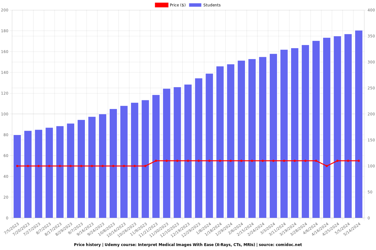
Price
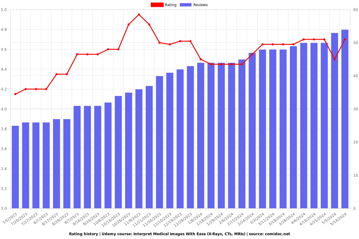
Rating
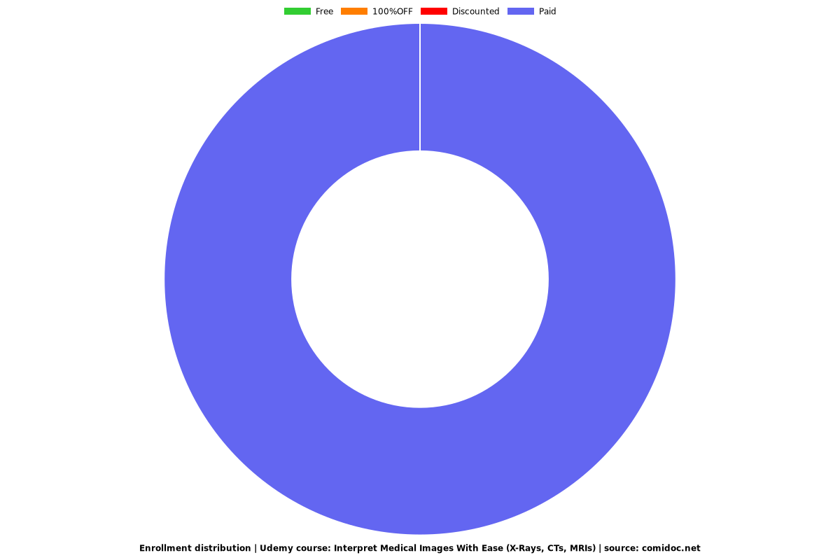
Enrollment distribution